


Informatics Educational Institutions & Programs

No higher resolution available.
Epithelial-cells.jpg (202 × 202 pixels, file size: 46 KB, MIME type: image/jpeg)
| Description |
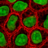
English: Cultured MDCK en:wikipedia:epithelial cells were stained for en:wikipedia:keratin, desmoplakin, and en:wikipedia:DNA. The stained cells were visualized by scanning laser confocal microscopy. The image shows how keratin cytoskeletal filaments are concentrated around the edge of the cells and merge into the desmoplakin which is located at en:wikipedia:desmosomes of the surface membrane. The network of keratin to desmosome to keratin linking the cells of an epithelial sheet is what holds together tissues like skin.
|
||||||||
| Source | wikibooks Cell Biology textbook (licensed under the GFDL): http://wikibooks.org/wiki/Image:Keratin.jpg | ||||||||
| Author | John Schmidt (user:JWSchmidt). | ||||||||
| Permission (Reusing this file) |
|

File history
Click on a date/time to view the file as it appeared at that time.
| Date/Time | Thumbnail | Dimensions | User | Comment | |
|---|---|---|---|---|---|
| current | 19:49, 2 May 2005 | | 202 × 202 (46 KB) | Helix84 | Cultured MDCK epithelial cells were stained for keratin, desmoplakin, and DNA. The stained cells were visualized by scanning laser confocal microscopy. The image shows how keratin [[Cytoskeleton|cytoskele |
File usage
The following pages on the English Wikipedia use this file (pages on other projects are not listed):
- Cell culture
- Cytokeratin
- Intermediate filament
- Metabolic engineering
- Tissue engineering
- User:Allthesestars/panel centre
- User:ClockworkSoul
- User:ClockworkSoul/userpage/panel center
- User:JWSchmidt
- User:JWSchmidt/Images
- User:Julie Zamostny/BIO134: Cancer Biology
- User:Lexor/Temp/Cell (biology)
- User:Mesoderm/sandbox
- User:Randoperson1/userboxes
- User:S spoerri/sandbox
- User:SnapJag/userpage/panel center
- Wikipedia:Facebook directory
- Wikipedia talk:WikiProject Molecular Biology/Molecular and Cell Biology/Discussion Archive
Global file usage
The following other wikis use this file:
- Usage on af.wikipedia.org
- Usage on anp.wikipedia.org
- Usage on ar.wikipedia.org
- Usage on az.wikipedia.org
- Usage on bg.wikipedia.org
- Usage on blk.wikipedia.org
- Usage on br.wikipedia.org
- Usage on bs.wikipedia.org
- Usage on ca.wikipedia.org
- Usage on ca.wikibooks.org
- Usage on cs.wikipedia.org
- Usage on cv.wikipedia.org
- Usage on da.wikipedia.org
- Usage on el.wikipedia.org
- Usage on en.wikibooks.org
- Usage on en.wiktionary.org
- Usage on es.wikipedia.org
- Usage on es.wiktionary.org
- Usage on et.wikipedia.org
- Usage on eu.wikipedia.org
- Usage on eu.wiktionary.org
- Usage on fa.wikipedia.org
- Usage on fi.wikipedia.org
- Usage on fr.wikipedia.org
View more global usage of this file.
